Perl's stain/Iron stain/Prussian Blue Reaction/Hematology/Special stain/STAR LABORATORY
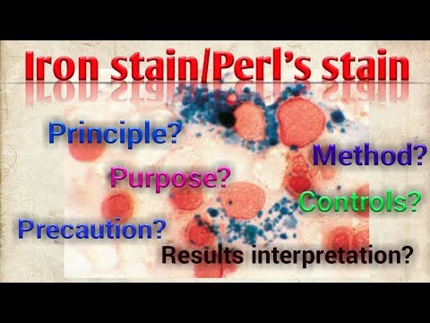
In this video you can watch the #practical on #Perl’s stain/ #Iron #stain. Iron stain is one of the special stain in #Histology and #Hematology. The #principle, method, purpose, controls, precautions, #results interpretations and other explanations are clearly given. Please leave your comments after watching this video, please let me know if there is any corrections.
Perls’ Prussian blue is a commonly used method in histology, histopathology, and clinical pathology to detect the presence of iron in tissue or cell samples.[1][2][3][4] Perls’ Prussian Blue derives its name from the German pathologist Max Perls (1843-1881), who described the technique in 1867.[2] The method does not involve the application of a dye, but rather causes the pigment Prussian blue to form directly within the tissue.[5] The method stains mostly iron in the ferric state which includes ferritin and hemosiderin, rather than iron in the ferrous state.
Perls’ method is used to indicate “non-heme” iron in tissues such as ferritin and hemosiderin,[6] the procedure does not stain iron that is bound to porphyrin forming heme such as hemoglobin and myoglobin.[2] The stain is an important histochemical stain used to demonstrate the distribution and amount of iron deposits in liver tissue, often in the form of a biopsy.[6][7] Perls’ procedure may be used to identify excess iron deposits such as hemosiderin deposits (hemosiderosis) and in conditions such as hereditary hemochromatosis.[8] Perls’ Prussian blue is commonly used on bone marrow aspirates to indicate levels of iron storage[4] and may provide reliable evidence of iron deficiency.
Perls did not publish a detailed procedure other than indicating a dilute potassium ferrocyanide solution was applied to the tissue followed by hydrochloric acid.[2] Ferric iron deposits in tissue (present mostly as ferric iron within the storage protein ferritin) then react with the soluble ferrocyanide in the stain to form the insoluble Prussian blue pigment (a complex hydrated ferric ferrocyanide substance). These deposits are then visualizable microscopically as blue or purple deposits.[9]
Many methods of performing Perls Prussian blue stain for iron have been published, [2] Drury and Wallington (1980) give a protocol that uses a mixture of 1 part 2% hydrochloric acid and 1 part 2% potassium ferrocyanide that is applied to the section for 20-30 minutes followed by a rinse in distilled water and application of a counterstain such as eosin, safranin or neutral red.
What is iron staining?
Hemosiderin staining. Hemosiderin — a protein compound that stores iron in your tissues — can accumulate under your skin. As a result, you may notice yellow, brown, or black staining or a bruiselike appearance. … When red blood cells break down, the hemoglobin releases iron.
What is Hemosiderin staining?
Hemosiderin staining is dark purple or rusty discoloration of the lower legs caused by chronic venous disease. A 2010 study found hemosiderin staining in all subjects with lipodermatosclerosis and venous ulcers. When vein valves fail, regurgitated blood forces red blood cells (RBCs) out of capillaries.
What is Prussian blue stain used for?
The Prussian blue Iron stain is used to demonstrate ferric (Fe3+) iron in tissues. The mode of action for the Prussian blue iron stain is to treat the tissue with an acidic solution (hydrochloric acid).
What is the Prussian blue test?
Perls’ Prussian blue is a commonly used method in histology, histopathology, and clinical pathology to detect the presence of iron in tissue or cell samples. … The method stains mostly iron in the ferric state which includes ferritin and hemosiderin, rather than iron in the ferrous state.
How does Perls stain work?
Perls Prussian Blue Staining Protocol. Dilute mineral acid hydrolysis releases ferric ions from protein bound tissue deposits, which, in the presence of ferrocyanide ions, is precipitated as the highly coloured and highly water-insoluble complex, potassium ferric ferrocyanide, Prussian blue
How does Prussian blue work?
How does Prussian blue work? Prussian blue traps radioactive cesium and thallium in the intestines and keeps them from being re-absorbed by the body. … Because Prussian blue reduces the time that radioactive cesium and thallium stay in the body, it helps limit the amount of time the body is exposed to radiation.
Comments are closed.